|
Obstetrics and Gynecological conditions of Cattle and Buffalo
Anoestrum
| Nature of disease |
|
- This is a metabolic disorder of dairy animals.
- This manifest digestive disturbance.
|
| |
Causes |
- Failure to detect / observe oestrus signs.
- Suboestrus, weak or silent oestrus.
- A low plane of nutrition, lack of energy and protein, deficiency of minerals namely P, Co, Fe, Cu, I, Mn and Vitamin A.
- Failure to recognize that an animal is pregnant.
- Anoestrus due to persistent corpus luteum, conditions associated with uterine pathology such as pyometra, mummified foetus, foetal maceration, mucometra and hydrometra and
- Insufficient hormonal stimuli.
- Senility, chronic debilitating disease like JD, TB etc., seasonal and environmental influences and heavy lactation (negative energy balance) may predispose anoestrus.
- Closely confined dark stables, lack of exercise combined with nutritive factors.
- In suckling animals prolactin may reduces the ovarian sensitivity
|
| |
Prevention Method |

TANUVAS Mineral Mixture |
- Unobserved oestrum may be due to managerial deficiencies and short period of oestrus. The dairy animals should be observed for heat signs at least three times a day.
- Wall charts, breeding wheels, herd monitors and individual cow records may be used for identify the oestrus.
- Teaser bulls (vasectomized or by applying apron) are useful in identifying heat in large number of animals especially buffalo cows.
- Provision of adequate lighting to improve oestrus detection.
- Silent / weak / Suboestrus are most common in buffalo cows and common in post partum period. In this cyclical changes in the genital organs occurs but the signs of heat are not exhibited or not observed. This requires rectal examination by qualified veterinary doctor.
- Extra feeding of a concentrate mixture or grains like maize, Cholam, kambu. Etc., and at least small amount of green fodder along with other roughages.
- TANUVAS Mineral mixture may be supplemented.
- After breeding the animals should be checked for pregnancy within 45-60 days by qualified veterinary doctor.
- Uterine pathology and hormonal stimuli should be handled by qualified veterinary doctor.
|
|
 |
Repeat breeder
| Nature of disease |
|
- Animals come to heat and not conceived after three successive inseminations are called repeat breeder.
- This may be due to various reasons
|
| |
Causes |
- Due to deficient Luteinizing Hormone release, delayed ovulation or failure of ovulation may leads to fertilization failure.
- Defective ovum or ageing of ovum may leads to fertilization failure.
- Inability of the sperm to fertilize a viable ovum.
- Inability of gametes to reach one another.
- The organisms Trichomonas fetus, Campylobacter fetus, Brucella abortus and IBR-IPV which may cause early embryonic death.
- Deficiency of Selenium and Vitamin E may cause early embryonic death.
- Long period of feeding estrogenic forages may affect the embryo survival.
- Environmental stress during first week after breeding may lead to early embryonic death.
|
| |
Clinical symptoms |
- Animal will not conceive even after three successive inseminations.
|
| Preventive and Control measures |
|
- Bring the animal into positive nutritive balance.
- Mineral mixture supplementation should be done to breeding animals.
- Do Artificial Insemination twice at each oestrus preferably at 12 or 24 hrs intervals.
- Skipping of AI and intrauterine infusions may be considered for uterine pathology.
- Diseased bulls should not be allowed for breeding.
- By avoiding diseased breeding bulls the pathogenic organisms causing abortion may be controlled.
|
|
|
Endometritis
| Nature of disease |
|
- Endometritis is a localized inflammation of the uterine lining, associated with chronic postpartum infection of the uterus with pathogenic bacteria.
|
| |
Causes |
- The causal organisms usually reach the uterus from the vagina at coitus, insemination, parturition or postpartum.
- The great majority of cows suffer from bacterial contamination of the uterus after calving.
- In cows that develop endometritis, the bacterial flora is not eliminated from the uterus, causing the endometrium to become inflamed.
- The factors associated with the development of endometritis are, retained fetal membranes, abortion, induced calving, multiple births, dystokia and bacterial loading.
|
| |
Clinical symptoms |
 |
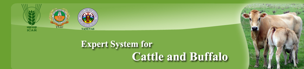
- The presence of a white or whitish – yellow mucopurulent vaginal discharge in the post partum cow.
- The volume of discharge is variable, but frequently increases at the time of oestrus when the cervix dilates and there is copious vaginal mucus.
- The cows rarely show any signs of systemic illness, although in a few cases milk yield and appetite may be slightly reduced.
- Rectal palpation frequently shows a poorly involved uterus which has a doughy feel.
|
| Control Method |
|
- A wide range of antibiotics, hormones, antiseptics and immunomodulators have been used as treatments for endometritis.
- This condition should be handled with qualified veterinary doctor.
|
|
|
Dystocia
| Nature of disease |
|
- The first or the second stages of parturition is markedly prolonged, becomes difficult or impossible for the dam to deliver the foetus without artificial aid.
- Dystocia means difficulty in birth.
- The overall incidence of dystocia varies with the species and with breeds within the species.
- The bovine species is most often affected.
- Dystocia is common in primipara than in pleuripara.
- Heavier male calves, twin pregnancy in cattle and low litter size in multiparous species, increase the incidence.
- Pregnancies that terminate early are conducive to dystocia through the medium of uterine inertia and fetal malposture.
- Prolonged gestation causing fetal oversize, close confinement, overfeeding, gross underfeeding and too early breeding increases the incidence.
|
| |
Causes |
- Twining, hydro amnion and foetal anasarca
- Improper nutrition of the growing heifers was the most important factor in retarding body and pelvic growth.
- Small pelvis, under developed juvenile genital tract, and lack of strength to expel the foetus.
- Breeding a poorly grown, underfed female that may be old enough to breed, but the body growth has been greatly retarded due to poor nutrition, parasitisms or diseases.
- High feeding levels favours excessive deposition of fat in the pelvic region predisposing to difficult parturition, especially in heifer.
- High feeding levels favours the development of a larger fetus (especially high feeding during the last third of pregnancy) leads to difficulty in giving birth.
- Close confinement of pregnant animals without exercise, are prone to torsion of uterus and uterine inertia.
- Any infection or disease affecting the pregnant uterus and its contents may cause dystocia.
- Dystocia may be due to Expulsive forces (Expulsive defect), adequacy of the birth canal (Constriction) and size and disposition of fetus (Over size and faulty disposition).
|
| |
Clinical symptoms |
 |
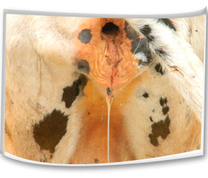
- Difficulty in giving birth.
- Calf limbs or face protrude from the vulva.
- Animal attempts strong forces and unable delivers calf
|
| Control Method |
|
- It should be handled with qualified veterinary doctor.
- It has been suggested that dairy heifers may be bred by size or weight rather than by age.
- During parturition all animals should be watched closely, if possible, so that prompt aid may be given if parturition is not normal.
- To help control infections that predispose to uterine disease and foetal death, both the sire and dam should be free of infection at the time of service.
|
 |
|
|
Total Uterine Prolapse
| Nature of disease |
|
- This is expulsion of uterine mass through the vagina.
- It is a common complication of third stage of labour.
- It is more common in pluriparous than primiparous animals.
- This condition is most commonly seen in cow than other animals.
|
| |
Causes |
- The flaccid atonic uterus.
- Violent or strong tenesmus during or after parturition.
- Retention of placenta at the ovarian pole of the uterine horn.
- Excessive relaxation of the pelvic and perineal region.
- Commonly seen in confined or stabled cattle especially during winter months.
- Forced extraction of the fetus.
- Over distension of the abdomen or excessive amounts of loose pelvic fat favour the condition by increasing the intra-pelvic pressure.
- Due to intra abdominal pressure.
- Delayed contraction of the uterus.
- Poorly grown, thin debilitated heifers and low plan of nutrition.
- During last 2-3 months of gestation, when large amounts of oestrogenic hormone being secreted by the placenta known as hyper estrogenism
|
| |
Clinical symptoms |

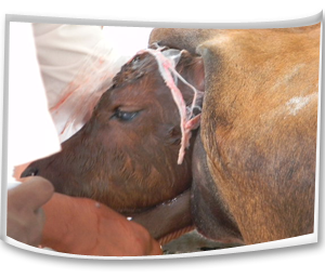
Hanging of uterine mass from the vulva |
- A mild protrusion of the vaginal mucous membrane through the vulval lips when the animal lies down.
- In standing posture, the prolapsed mass hanging upto the hock joint.
- The fetal membranes and /or mucus membrane of the uterus is exposed.
- The mass is usually covered with feces, straw, dirt or blood clots.
- Uterus is enlarged and edematous especially in delayed cases (4-6 hrs).
- The cervix is usually present at the vulva.
- The non gravid horn is held inside the peritoneal surfaces of the prolapsed gravid horn and does not evert.
- Stress, restlessness, pain, anxiety, increased respiratory rate may be noticed.
- Internal hemorrhage due to rupture of one of the uterine vessels, shock, incarceration of the intestinal mass and death.
- Pale mucus membrane, expiratory grunt and prostration with severe depression and inability to rise indicated serious complications.
|
Protrusion of vaginal mucous membrane |
| Suggested first aid |
|
- The prolapsed animal should be separated from other animals.
- The mass should be protected from contamination from out sources.
- The mass should be covered with wet clean clothes until treatment.
- It needs careful insertion of protruded uterine mass into the body and suturing the in the vulval lips so this condition should be handled with qualified veterinary doctor.
|
|
|
Torsion of Uterus
| Nature of disease |
|
- Uterine torsion is defined as the twisting or revolution of the gravid uterus on its long axis and is common in cows and buffaloes.
- High incidence among pluriparous than primiparous
|
| |
Causes |
- Frequent lying down and getting up may predispose to uterine torsion.
- Lack of fetal fluids and violent falling or rolling and sudden movements may predispose to torsion.
- Close confinement of pregnant animals for a longer period may favour occurrence of uterine torsion.
- A lack of tone of the pregnant uterus comprising lack of fluids, flaccid uterine walls, a small non gravid horn, a long flaccid mesometrium favours uterine torsion.
- A deep capacious abdomen predispose to uterine torsion and especially in buffaloes because of the wallowing habit.
- Exiting causes such as horn thrust or butting by the neighboring animals, violent movements during grazing, rolling due to tympany and colic may cause torsion.
- Transportation of pregnant animals from one place to another in rail or by road cause violent and irregular movements creating anxiety and predispose to torsion of uterus.
|
| |
Clinical symptoms |
- The signs of abdominal pain, anorexia, constipation, lack of ruminations, restlessness or colic symptoms, teeth grinding, treading and tail switching may be observed.
- Displacement of dorsal commissure.
- Tucked up udder and
- Vulval edema.
|
| Prevention Method |
|
- Pregnant animals should be housed separately and extra care should be given in handling the animals.
- Rolling, jumping, frequent lie down and falling of pregnant animals should be avoided.
- Transportation of pregnant animals should be avoided.
- Attack and fight with other animals should be avoided.
|
| |
Control Method |
- This condition must be handled with qualified veterinary doctor.
- Rotation/ rolling of the dam may be performed with the help of veterinary doctor.
- In complicated cases cesarean or Laparo- hysterotomy may be recommended.
|
|
|
Retained Fetal Membranes
| Nature of disease |
|
- This is one of the most common conditions occurring in animals following parturition.
- This is failure of the villi of the fetal cotyledon to detach from the maternal crypts of the caruncles and retained longer than normal time limits.
- If the placenta is retained longer than 8-12 hours the condition is considered as pathological.
|
| |
Causes |
- Infections of uterus during gestation may be a cause for retained placental membranes.
- Uterine inertia due to hormonal imbalance such as low level of oxytocin.
- Deficiency of Vitamin A and iodine causes retained placenta.
- The hormone progesterone and excess of cortisol in late gestation may cause the retension of fetal membranes.
- The disease conditions causing uterine inertia or atony results in a higher incidence of retention of foetal membranes.
- Close confinement and lack of exercise highly prone to retained placenta.
|
| |
Clinical symptoms |
- A portion of the fetal membranes hang from the vulva 12 hours or more even after the expulsion of the foetus.
- Anorexia and depression may develop.
- A fetid odour develops since the placenta begins to macerate after 24 hours of foetal expulsion.
|
| |
Control Method |
- It should be handled with qualified veterinary doctor by manual removal.
- Before arriving veterinarian, the hanging foetal membranes should be protect from dogs or other animals.
- The protruding membranes should be tied in a knot to prevent them touching the hocks.
- Manual removal can be attempted in 24- 48 hours after parturition.
- Manual removal after 48 hours is not advisable because cervix was closed.
- Manual removal of placenta is contraindicated in cows with elevated temperature and also with vaginitis and vulvitis.
- After removing the fetal membranes, tetanus toxoid injection is recommended to prevent tetanus infection.
|
|
|
|